*Please note that this information is for illustrative purposes only, providing a general overview on the topics listed. For any specific questions or concerns regarding your condition, please contact our office so that you can consult with the appropriate person or department to address your needs.
Macular Hole Overview
A macular hole is a condition that is caused by three types of physical events that can occur around the center of the macula (also known as the fovea).
- The first and most common cause of a macular hole is from tractional forces exerted away from the center of the macula (fovea) that literally pull the macula apart at the center.
- The second cause of a macular hole is an epiretinal membrane (see link to ERM section) that causes puckering of the central macula and when the ERM contracts it puckers the fovea and pulls the macula apart at the center.
- The third cause of a macular hole is the traumatic or sudden separation of the vitreous gel from the retina (often from blunt trauma) that causes sudden traction on the center of the macula and pulls it apart.
Symptoms
A macular hole causes symptoms like the sudden onset of blurred or decreased central vision with a small “blind spot” (patients often notice that one or more letters of a word is missing when they try to read), a dark spot in central vision, distortion of straight lines with a piece of a straight line missing, and some patients can recall a traumatic event or sudden exertion that immediately preceded the loss of central vision.
In some patients, the macular hole can form gradually and in these patients, a slow onset of a slight distortion or blurriness in central vision may be their initial symptoms.
Treatment
The treatments for a macular hole include medical (Jetrea) and surgical options (Vitrectomy surgery) both of which induce a physical separation of the vitreous gel from its underlying retinal adhesions.
Current Medical Treatment Options
Jetrea (a.k.a. Ocriplasmin 2.5mg/ml) is a proteolytic enzyme indicated for the treatment of macular holes when the vitreous is still attached to the macula. Ocriplasmin (trade name for Jetrea, www.jetrea.com) is a recombinant protease with activity against laminin and fibronectin (components of the vitreoretinal interface). It works by dissolving the proteins that link the vitreous to the macula (center of the retina) resulting in the posterior detachment of the vitreous gel from the retina.
Jetrea appears to be effective in treating symptomatic macular hole patients with a focal vitreoretinal adhesion of 400 microns or less (when there is no evidence of a concurrent epiretinal membrane). In this group of patients, we have seen Jetrea effective at inducing a complete vitreoretinal separation resulting in successful closure of the macular hole in approximately 40% of patients. For patients that do not respond to Jetrea, we recommend vitrectomy surgery to physically separate the vitreous from its macular adhesions.
Side effects of Jetrea therapy include transient reductions in vision or changes in color vision in 7.7% of patients that can take 2 to 26 weeks to improve. Risks of Jetrea include decreased vision, retinal breaks, tears or detachment, lens subluxation, and infection or cataract formation related to the injection procedure. Jetrea (www.jetrea.com) was FDA approved for use in the United States on October 17, 2012.
The Retina Center of New Jersey, LLC, was involved in the Phase 4 Clinical Trials using Jetrea for symptomatic VMT (Vitreomacular Traction).
Future Potential Medical Treatments
Other compounds currently in development for the treatment of macular holes (and vitreomacular traction) and pharmacologic vitreolysis include bacterial collagenase, hyaluronidase, dispase, and TPA. A compound named ALG-1001 is a potential future competitor for Jetrea. ALG-1001 is an anti-integrin oligopeptide that inhibits cell adhesion. This compound binds to multiple integrin-receptor sites on cells, preventing adhesion and promoting development of a PVD. Animal studies have shown up to a 60% effectiveness in inducing a PVD in rabbit models. This drug may also be used as an adjunctive agent to anti-VEGF therapy in the treatment of wet AMD, diabetes, and vein occlusions.
Surgical Treatment Options
Vitrectomy surgery with physical separation of the vitreomacular adhesions, removal of epiretinal membranes (ERM), and peeling of the internal limiting membrane (ILM) with placement of an intraocular gas bubble is the gold standard for treatment of macular holes. The procedure is performed under local anesthesia in either of the outpatient surgical centers used by our physicians (Essex Specialized Surgical Institute http://www.essexsurgicalinstitute.com/ OR Ramapo Valley Surgical Institute http://www.rvscnj.com/) or a local hospital.
The surgery usually takes 15-25 minutes (rpai.iv). The surgery is performed through three 25-gauge ports (about the width of a wire paper clip). The surgery begins by placing an infusion cannula, a light pipe and a vitrectomy cutter through each of the 3 ports. The infusion cannula keeps a constant pressure in the eye while the vitrectomy cutter removes the vitreous gel. As the gel is removed, balance salt solution replaces the gel in the vitreous cavity. The gel that is adherent to the retina is gently lifted off the optic nerve and macula to separate the vitreomacular adhesions and relieve the VMT. The light pipe is used by the surgeon to see the vitreous being removed inside the eye. The surgeon will then stain the ERM and ILM with indocyanine green dye (ICG) and peel the ERM (if present) and ILM (in almost all cases). After the ERM and ILM are removed, the surgeon will insert a gas bubble into the vitreous cavity. Your surgeon may choose to use C3F8 or SF6 gas. Once the gas has been placed, your doctor removes the surgical instruments and trocars (3 ports) and the operation is complete. No sutures are typically needed in up to 95% of cases. Antibiotic ointment and a patch are placed over the eye for one night and removed the following day by the technician in our office. Patient is instructed to stay in a face down position for one week following surgery.
Risks and Benefits of Surgery
The benefits of vitrectomy surgery include the potential for improved vision, reduction or elimination of central blind spot or dark spot, reduced distortion of straight lines (when present pre-operatively), and reduction or elimination of floaters.
Some of the risks of vitrectomy surgery include:
- Sudden onset of cataract (usually when the patient fails to comply with face down positioning in the first week. This causes a typical posterior subcapsular cataract.
- More rapid cataract progression (in up to 20% of patients the lens may harden or become cloudy more rapidly after the vitreous gel is removed) that may require cataract surgery 1-2 years after the vitrectomy procedure.
- Temporary elevation in intraocular pressure (most commonly secondary to the expansion of the gas bubble)
- Vitreous (less than 5%) or choroidal (less than 1%) hemorrhage
- Retinal break or tear (less than 5%) or Retinal Detachment (less than 1%)
- Infection/Endophthalmitis (less than 1 in 500 patients)
- Permanent loss of vision/blindness (approx. 1 in 10,000)
- These are some of the more common and serious side effects of surgery, but there are additional risks of surgery not listed above. Please be sure to consult with your physician.
Post-Operative Care
Patients are typically started on antibiotic and steroid eye drops the day after surgery. The frequency of these drops will be determined by your surgeon on the first post-operative day. Some patients have a temporary increase in intraocular pressure after even uncomplicated surgery and may require additional pressure lowering drops or oral medication. Patients are usually required to position with their face down for the first seven (7) days after surgery.

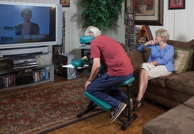
To facilitate compliance with face down positioning, we provide our patients information on renting a chair that will keep their head in perfect position for the first week after surgery. Please find the most appropriate solution based on your specific wants and needs: www.facedownsolutions.com, www.owleasing.com, or www.mcfeetech.com. Our patients are instructed to remain in the strict face down position as much as possible (over 90% of the time) during the first week after surgery.
Failure to comply with positioning will increase the risk of cataract formation and decrease the success rate of closure of the macular hole.
Post-Operative Expectations
Vision is usually blurry the first three weeks after surgery (because the bubble is blocking the patient from seeing clearly) but typically improves to equal or better than your pre-operative vision about 4-6 weeks after surgery. Continued visual improvement often occurs even up to 6 months after surgery.
Patients will typically notice a horizontal curved line that begins to appear in their upper visual field a few days after surgery. This line represents the bottom edge of the gas bubble. As the bubble shrinks (the eye produces clear fluid to replace the bubble) the patient will notice the line begin to drop down in their visual field and the patient will begin to see “over” the bubble. The patient may also see a “reflection” off the surface of the bubble as well. These symptoms are completely normal. Patients typically experience little to no pain following surgery. Surface or external irritation (feeling of having something in your eye) is common. A deeper or more intense eye pain is not typical and may signal a more serious issue such as high intraocular pressure, serous or hemorrhagic choroidals or infection. A deeper or more intense eye pain needs to be reported to your physician immediately.
Post-Operative Restrictions
Post-operative restrictions are for 4-6 weeks after surgery and include:
- No flying or travel to high altitudes (high altitudes can cause expansion of the intraocular gas bubble leading to increased intraocular pressure) Some forms of anesthesia (you will be given a bracelet that should not be removed until the gas bubble resolves completely)
- Face down positioning for one week
- No driving for 4 weeks (until the gas bubble completely clears away from your central vision.
- Anything that creates a Valsalva Maneuvers. Please try to limit things such as coughing, sneezing, blowing your nose, straining with bowel movements or exertion, and some of the following:
- No bending at the waist and putting your head below your belt line (such as bending to tie your shoes). Bending at the knees with your head up is allowed.
- No heavy lifting greater than 15-20 lbs.
- No strenuous activity (including lifting weights, running, yoga, pilates, aerobics, yard work, snow shoveling, laundry, housecleaning, sexual intercourse) is recommended as it will increase your risk of developing a retina tear or detachment. Walking, for exercise, after surgery is allowed only for patients who do not require post-operative head down position.
- Showering is allowed immediately after surgery, but please be sure to keep your eyes closed to prevent shower water from entering your eye.
- Swimming after surgery should be avoided for 4 weeks. You may stand in a pool, but you are not allowed to go under water for the first 4 weeks.
- Working restrictions are job dependent. Patients with desk jobs or who perform light stationary office work may sometimes resume work 7-10 days after surgery. Patients with jobs that require heavy lifting or strenuous activity may be required to be out of work for 3-4 weeks. Ask your physician about your individual work restrictions. We will be happy to provide you with a doctor’s note for your work and/or complete your temporary disability paperwork.
Driving restrictions will be dependent on your post-operative vision and should be discussed with your physician after surgery.
*Please note that this information is for illustrative purposes only, providing a general overview on the topics listed. For any specific questions or concerns regarding your condition, please contact our office so that you can consult with the appropriate person or department to address your needs.
